Health Care Provider Dental Health Issues
A recent case was completed in our office. A 53-year-old male health care provider presented with upper anterior teeth in the "esthetic zone" that were failing; the teeth were bridged together, but were becoming mobile partly due to decay and breakdown and to periodontal involvement as well. Much of the patient's problems were due to years of neglect. The image below reveals the patient prior to the beginning of care.
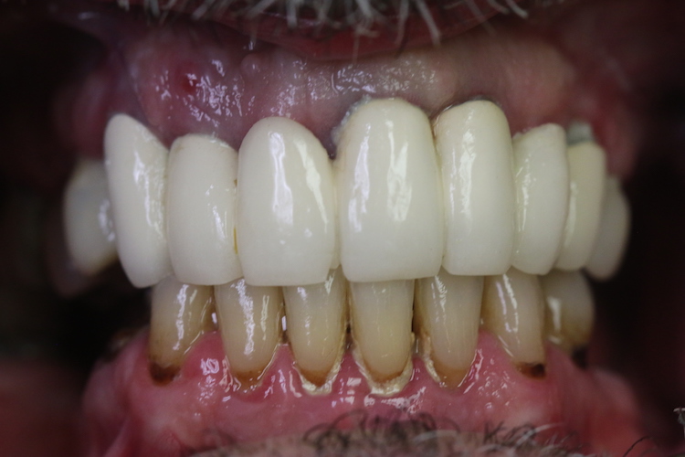
The patient prior to the beginning of care
After a complete clinical and radiographic review, a treatment plan was developed. Upon the patient's acceptance and consent the case was begun. Preliminary impressions were taken and the state of the dentition was documented with intraoral photography.
The upper anterior teeth were extracted and a laboratory pre-prepared Provisional Partial Upper Denture was placed to restore the dentition to form and function until the planned care was completed. Images of the Provisional Partial Upper Denture found below was worn intraorally until the completion of care.
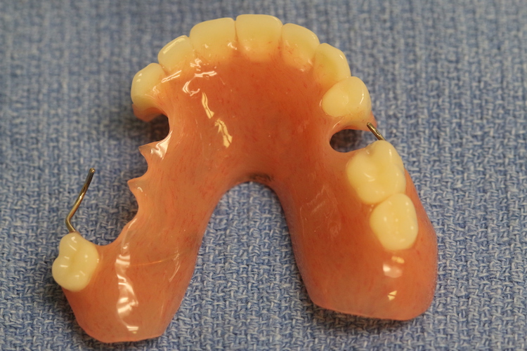
The Provisional Partial Upper Denture (1 of 2)
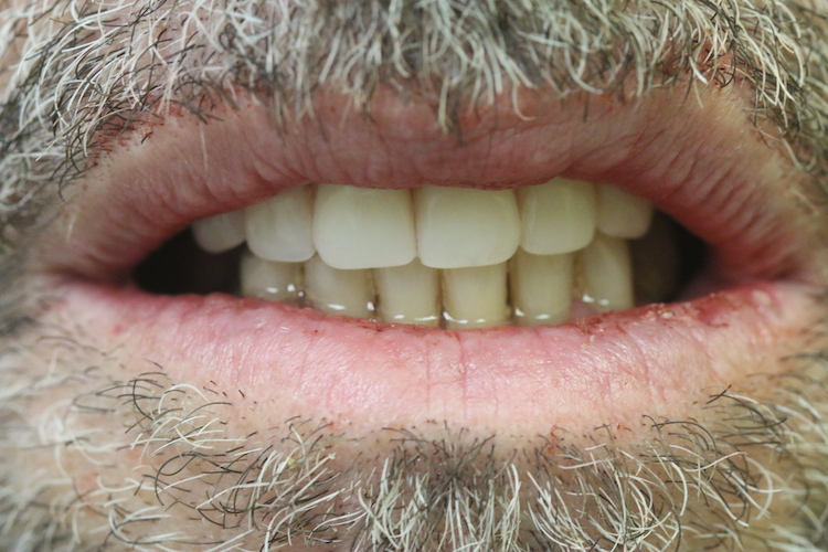
The Provisional Partial Upper Denture (2 of 2)
After sufficient healing time the upper anterior residual bone ridge was accessed by splitting the residual soft tissues open, and bone grafting the areas where implants were planned to be placed. An image of the soft tissues is below.
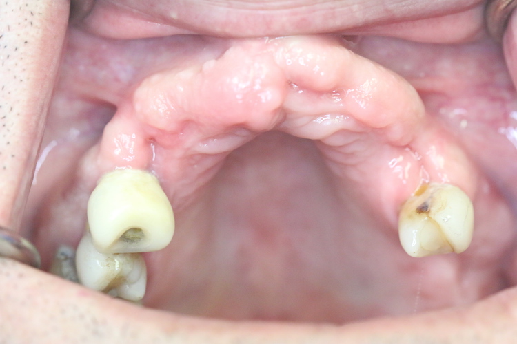
Soft tissue in the patient
Following 5 months of healing the patient underwent a Cat Scan to evaluate the bone and provided a means of measuring the bone in three dimensions. It also digitally provided a means of fabricating a template to place the planned implants at specific locations at measured distances and angulations so that the planned bridge that would be supported by the implants would restore form and function to the dentition. An image of the template in place intraorally is found below.
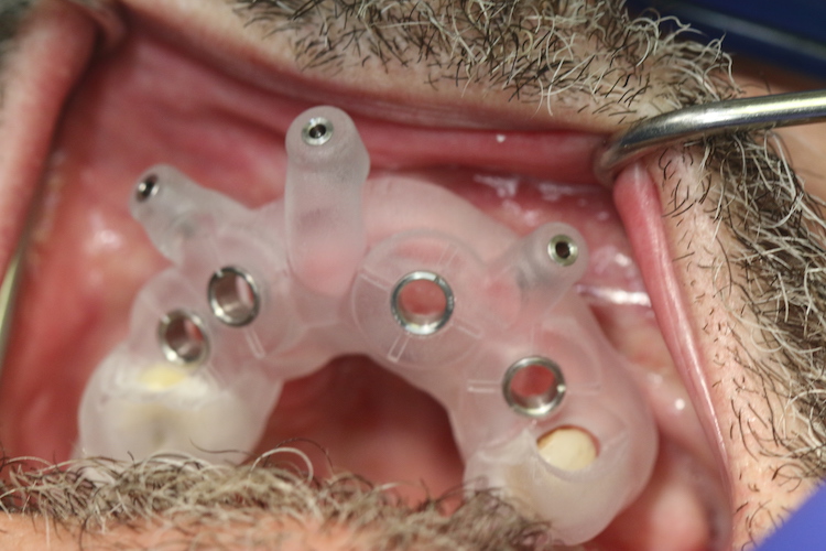
The template in place intraorally
With the template in place the projected implant sites were prepared and the implants placed correctly spaced and at the ideal angulation. The implants are allowed to heal for about 4 months and then an impression is taken to capture the spacing and angulation. An image below reveals the implants following four months of healing and with the metal abutments in place prior to placing the planned implant supported bridge.
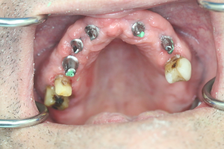
The implants following four months of healing and with the metal abutments in place
The images below are that of the completed case.
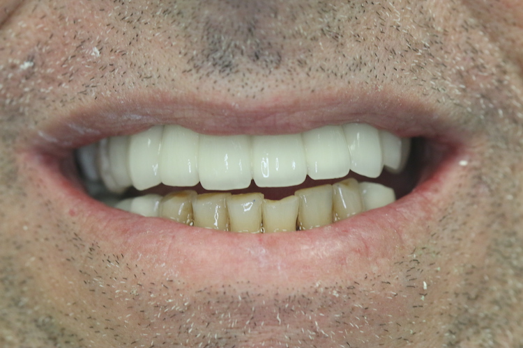
The completed case (1 of 3)
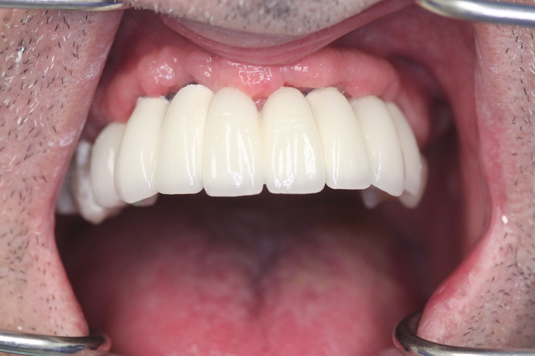
The completed case (2 of 3)
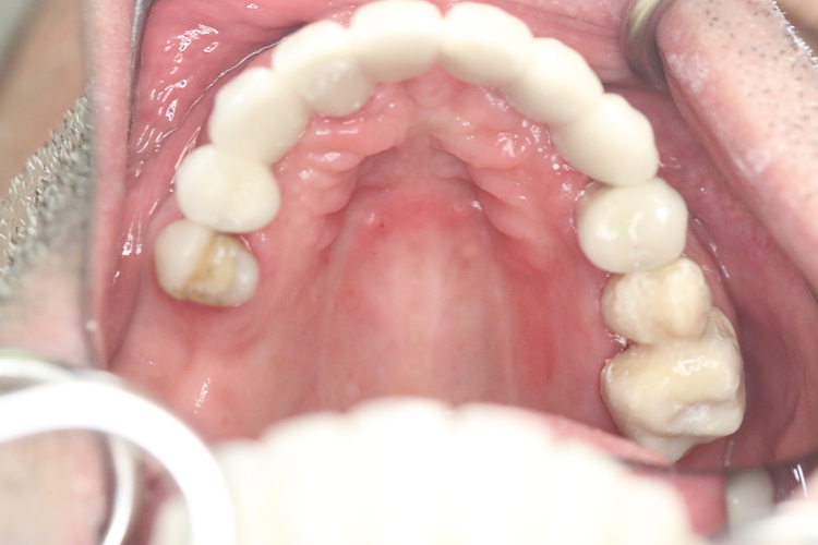
The completed case (3 of 3)
If you would like to discuss this case or any other at the eastboyntondental.com website, please contact us at (561)734-8600.


